Core Facility Campus Krems
Facility
Imaging
Electrophysiology Setup for Tissue Slices
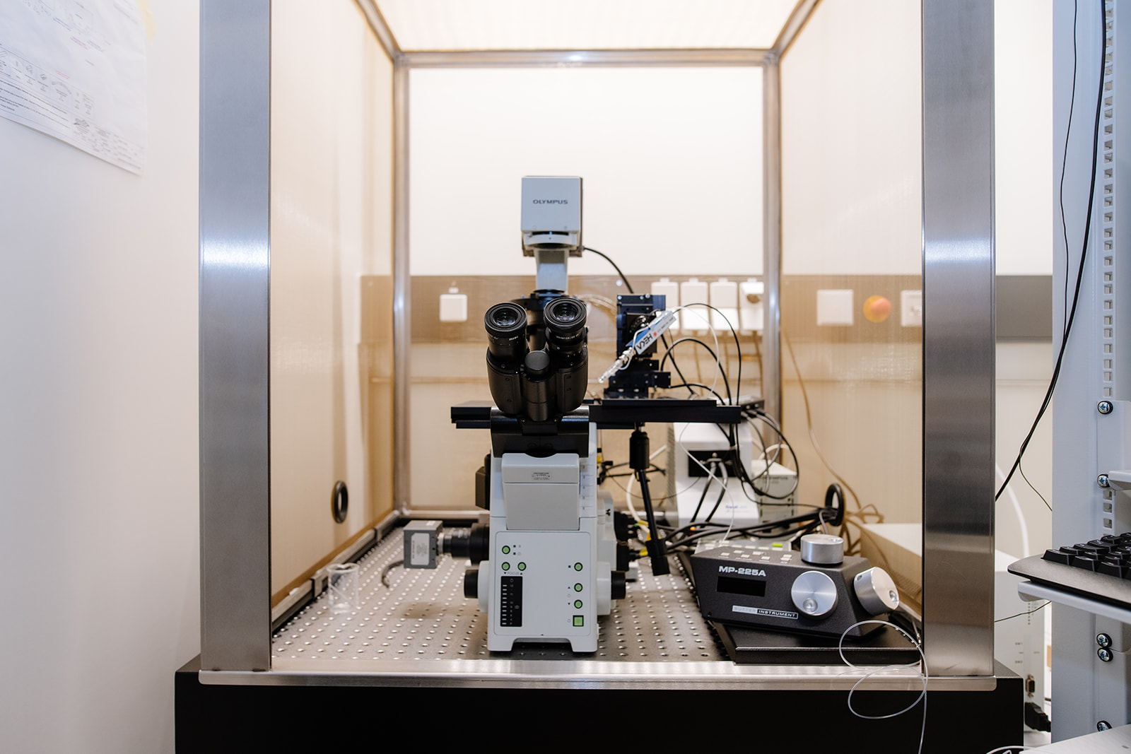
This electrophysiology setup is designed for in vitro studies on freshly prepared (alive) tissue slices, with a primary focus on mouse brain tissue. It consists of an upright fluorescent microscope (Olympus BX51) and an infrared camera, facilitating high-resolution visualization of cells located within deeper regions of the slice. The setup enables the measurement of endogenous ionic currents and spontaneous action potentials in nerve cells, thus allowing the study of neuronal activity in a physiologically relevant context.
In addition, combining electrophysiological recording with simultaneous, localized stimulation of adjacent brain slice regions with special electrodes or biochemically active compounds, allows for the study of synaptic transmission within complex neuronal brain circuits. Thus, the impact of prospective pharmacological agents on neuronal activity and signal transmission can be examined utilizing this experimental setup.
Location: Karl Landsteiner University of Health Sciences
Contact person: Prof. Dr. Gerald Obermair
Electrophysiology setup / patch clamp Olympus
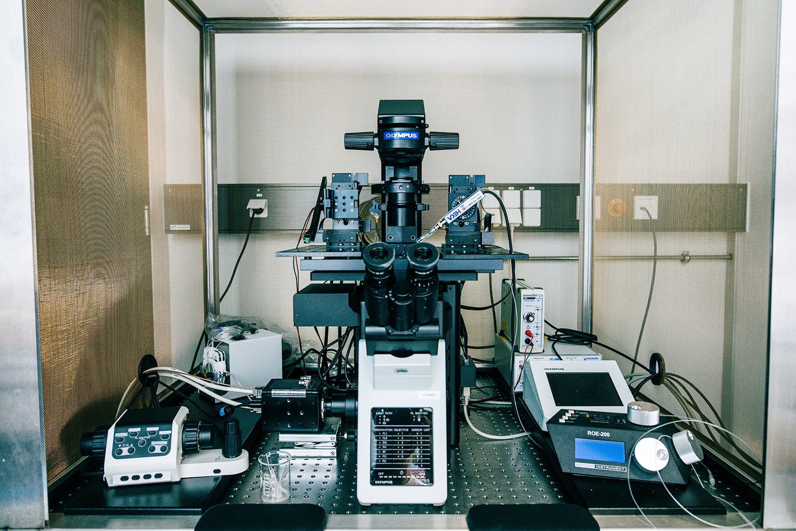
The electrophysiology setup consists of an inverted research microscope for high-resolution in combination with a 2-channel electrophysiology system, which allows intracellular electrophysiological measurements under simultaneous microscopic control. The Setup is primarily used for analysing two different samples:
- the analysis of cultured cells allows the recombinant expression of ion channels and their regulatory subunits. The channels expressed in this way can be measured and analysed with regard to their functional properties using the electrophysiology setup. This mainly serves to analyse the biophysical properties of the channels and thus allows predictions to be made about the physiological functions and the development of possible agonists or antagonists.
- the analysis of mouse brain slices: The setup also allows the measurement of neuronal connections and interactions in acute (living) mouse brain slices. This allows the communication of nerve cells in the existing neuronal system to be analysed and thus statements to be made about the function of ion channels for physiology and pathophysiology (e.g. the development of neuropsychiatric diseases).
Location: Karl Landsteiner University of Health Sciences
Contact person: Prof. Dr. Gerald Obermair
Epifluorescence microscope / Olympus
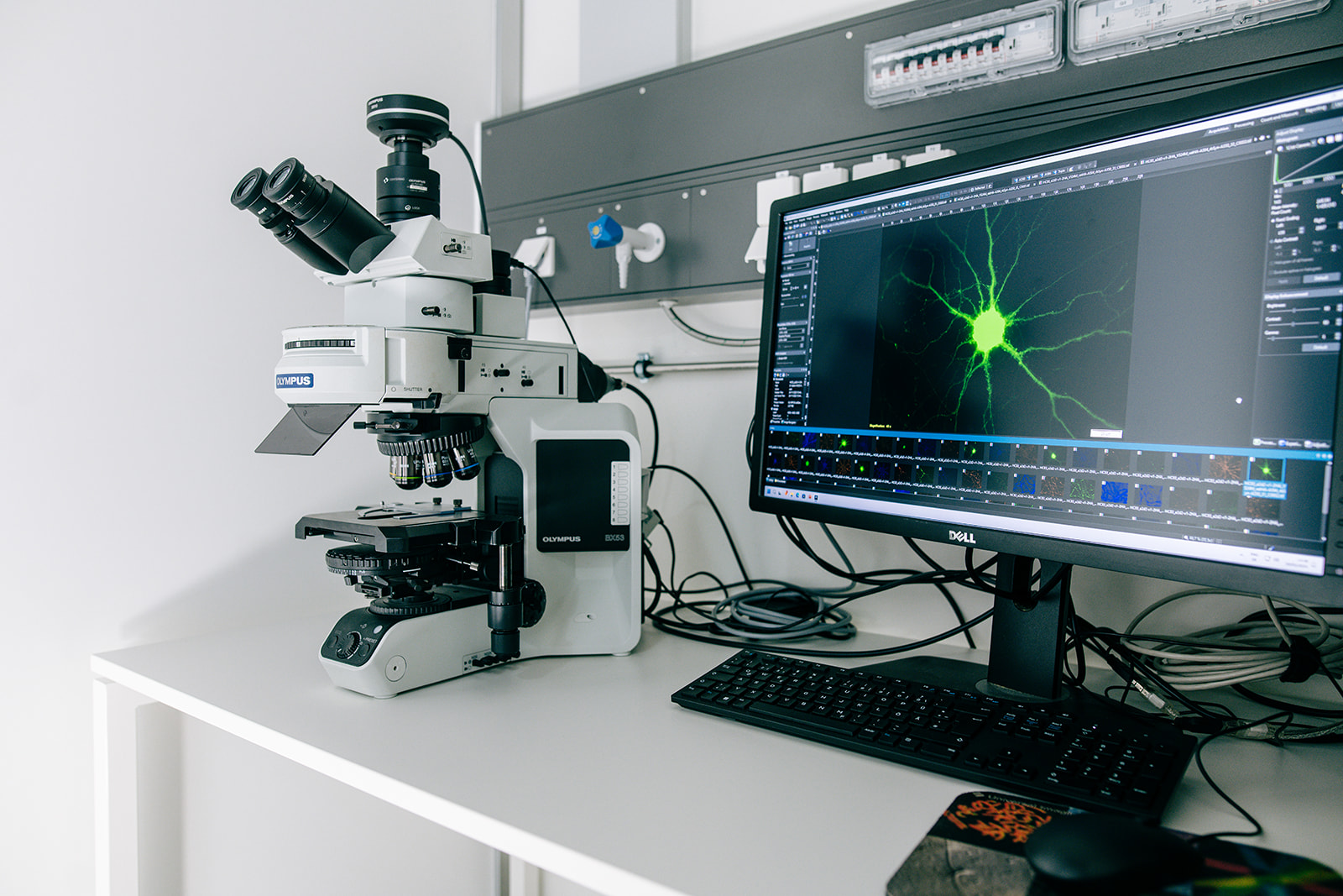
This Olympus BX53 microscope is equipped with Lumencor Sola LED light source, 14 bit CCD camera (XM10), objectives, control software and monitor.
Location: Karl Landsteiner University of Health Sciences
Contact person: Prof. Dr. Gerald Obermair
Confocal Laser Scanning Microscope
TCS SP8 AOBS
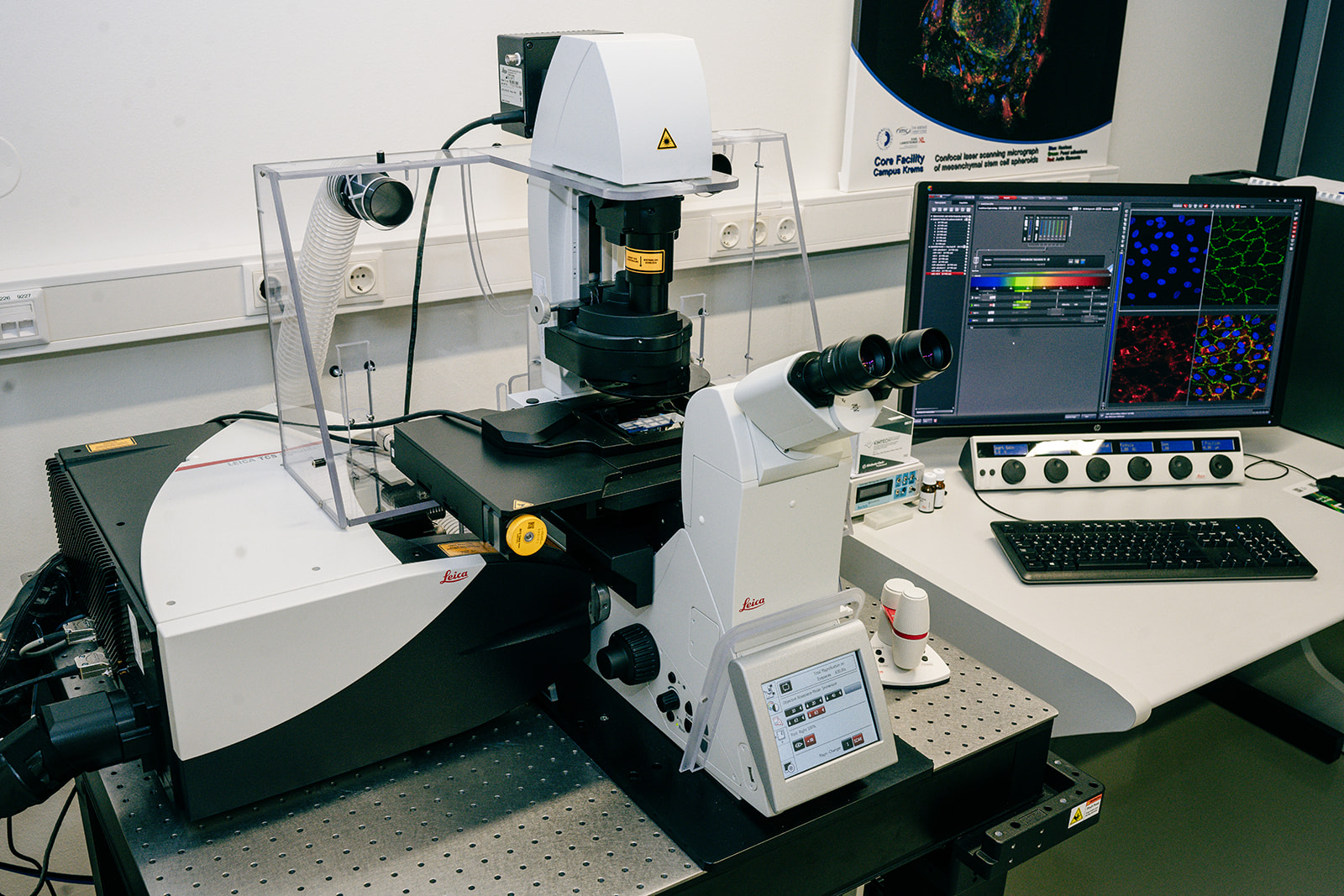
The Leica TCS SP8 is a confocal laser scanning microscope optimized for high-resolution and fast live-cell imaging, as well as high-content screening. By employing laser light and a pinhole aperture to reduce out-of-focus light, confocal laser scanning microscopy enables detailed observation of fluorescently labelled structures of biological and material specimens via optical sectioning. An incubation chamber provides optimal conditions for imaging living cells and to monitor dynamic processes. The Leica HyD detectors provide super-sensitive photon detection and are ideal for imaging living cells as well as samples with low fluorescence intensities and provide an improved signal-to-noise ratio.
Technical Details
- Inverse microscope DMi8
- TCS SP8 confocal scanner with Acoutso-Optical Beam Splitter (AOBS) and resonant scanner 8 kHz
- Hardware auto-focus control (AFC)
- Detectors: 2 GaAsP-Hybrid Detectors (HyDs), 2 Photomultiplier Tubes (PMT), 1 Transmitted Light Detector (TLD)
Lasers and Laser Lines
- Diode 405 (405 nm)
- Argon (458 nm, 476 nm, 488 nm, 496 nm, 514 nm)
- DPSS 561 (561 nm)
- HeNe 633 (633 nm)
Objectives
- HC PL FLUOTAR 10x/0.30 DRY
- HC PL APO CS2 20x/0.75 IMM
- HC PL APO CS2 40x/1.10 WATER
- HC PL APO CS2 63x/1.30 GLYC
- HC PL APO CS2 63x/1.40 OIL
LAS X Software Modules
- LAS X SP8 Software License for 2D- and 3D-image analysis
- 3D Visualization Module for reconstruction and data processing
- Live Data Mode Module for live-cell experiments
- HyVolution2 Module for confocal super-resolution
- Software Module for mosaic scans and multi-position experiments
- Navigator Module to create overviews, identify areas of interest (ROIs), and set up automatic high-resolution image acquisition
- Dye Finder
Location: University for Continuing Education Krems / D.2.20
Contact person: Tanja Eichhorn, MSc PhD
Nanoparticle Tracking Analysis (NTA)
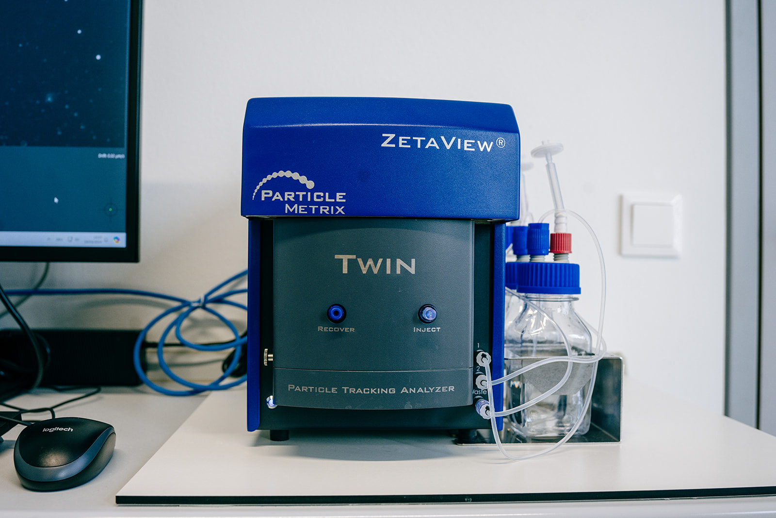
The ZetaView PMX-230-Z TWIN Laser System from Particle Metrix is a nanoparticle tracking analyzer, that measures the size distribution, concentration, as well as the zeta potential of particles in suspension. The instrument is equipped with a blue (488 nm) and a red (640 nm) laser to enable the detection of fluorescently labelled biomarkers. Due to the linked fluorescent modes, the instrument is suitable for co-localization experiments on double-stained samples. This device can be applied for the physicochemical as well as phenotypical characterization of extracellular vesicles and nanoparticles.
Location: University for Continuing Education Krems / D.2.23
Contact person: Vladislav Semak, PhD
Scanning Electron Microscope
Videko Hitachi Flex SEM 1000

Compact VP-REM electron microscope plus control PC. The device is a benchtop or compact SEM for imaging-oriented tasks that go beyond the performance spectrum of conventional benchtop SEMs. It offers excellent imaging performance and detector variability in compact dimensions.
Technical features:
- Convenient sample navigation
- 3-axis automation, manual Z-axis and tilting
- High-resolution electron optics up to 4 nm at 20 kV
- High ease of use
- Innovative navigation function
- Compact design
Fully-fledged, easy-to-operate device with compact size
Location: University for Continuing Education Krems / D.2.18
Contact person: Assoz. Prof. Dr Jens Hartmann