Core Facility Campus Krems
Facility
Molecular Biology and
Biochemistry
ChainRotary Cell Culturing System (RCCS)
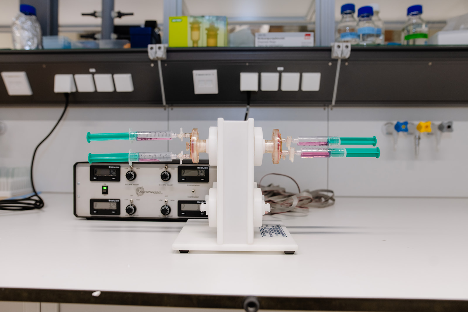
The Rotary Cell Culture System (RCCS) is a bioreactor for three-dimensional cell culture under conditions that approximate the in vivo environment. Cells are maintained in continuous suspension within a rotating vessel, creating a low-shear, low-turbulence environment that promotes spheroid formation and growth. Efficient nutrient and oxygen transfer reduces necrosis in the spheroid core, enabling long-term culture of complex aggregates.
The RCCS supports co-culture of multiple cell types and can be used with or without scaffolds to generate 3D models. The RCCS-4D configuration operates up to four vessels at the same adjustable rotation speed.
Typical applications include stem cell maintenance and differentiation, cancer spheroid modeling and invasion studies, tissue engineering, organoid culture, and host-pathogen interaction research. The system provides a versatile platform for developing physiologically relevant 3D cell culture models.
Location: Karl Landsteiner University of Health Sciences
Contact person: Prof. Dr. Klaus Podar
Digital PCR
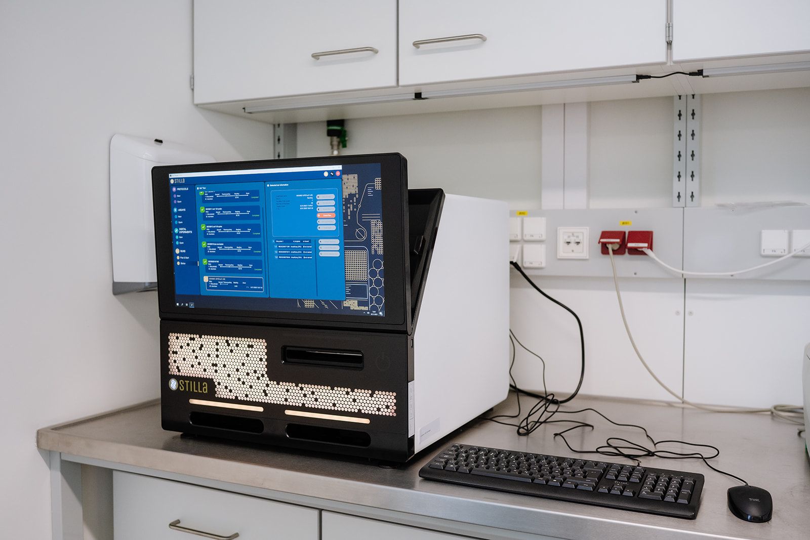
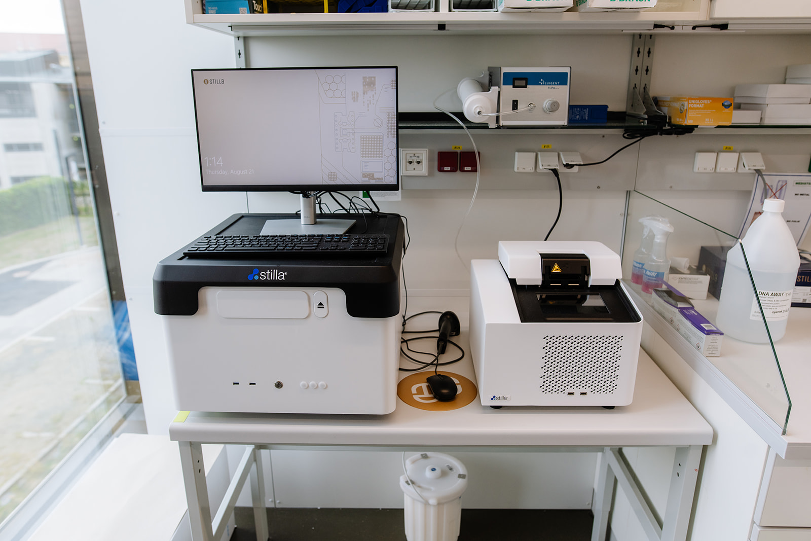
Digital PCR (dPCR) is a highly precise approach for the highly sensitive detection and quantification of nucleic acids. Compared to conventional real-time qPCR, dPCR does not require standard curves for quantification, as each sample is divided into thousands of individual reactions. This allows the absolute number of molecules present in the sample to be calculated. Further advantages include high tolerance to PCR inhibitors and the ability to detect mutations and validate standards for conventional qPCR assays.
Location: Karl Landsteiner University of Health Sciences / KL Y 2B-010
Contact persons: Assoc. Prof. Dr. Alexander Kirschner ; Dr. Claudia Kolm
Chemidoc MP / Bio-Rad, upgrade kit for fluorescence
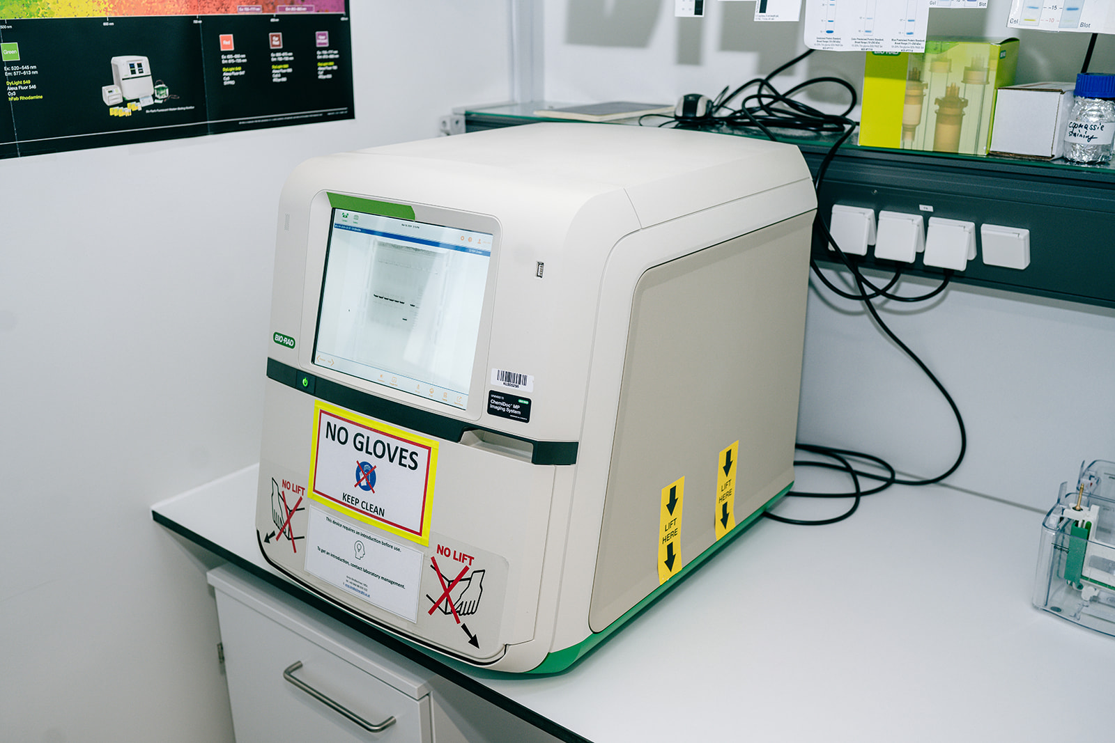
Gel imagers are essential devices in every molecular biology and medical laboratory and enable genetic material and protein samples to be analysed qualitatively and quantitatively. This can provide crucial insights into the molecular biological processes within a cell and is used worldwide as a robust method for detecting protein expression and analysing genetic traits. The research location Krems is home to a large number of research groups with a molecular biology focus that benefit from such a device.
Location: Karl Landsteiner University of Health Sciences
Contact person: Prof. Dr. Gerald Obermair and Prof. Dr. Dagmar Stoiber-Sakaguchi
Optima MAX BioSafe Ultracentrifuge / Beckman Coulter
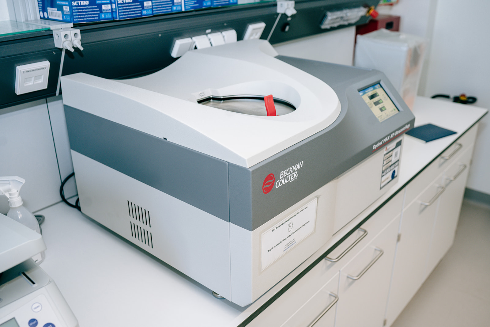
Ultracentrifuges are devices that generate gravitational fields through high rotational speeds and can thus cause solid components of a solution to sediment. This can be used to pelletise cells, nucleic acids or other solid components, but also to create density gradients. Ultracentrifuges are widely used in sample preparation and are essential for molecular biology laboratories.
Location: Karl Landsteiner University of Health Sciences
Contact person: Prof. Dr. Gerald Obermair and Prof. Dr. Dagmar Stoiber-Sakaguchi
qPCR Light Cycler 96 / Roche
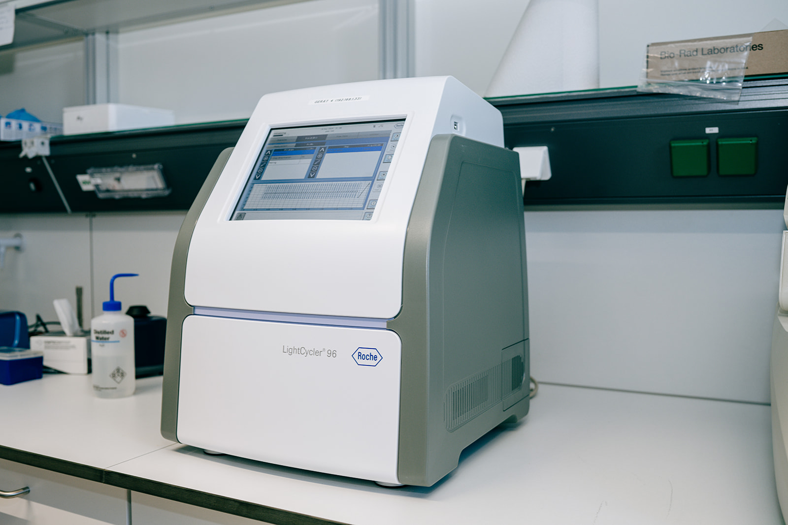
Real-time PCR (RT-PCR) devices enable the amplification and detection of very small amounts of DNA and can be used to quantify gene expression patterns, among other things. RT-PCR is a globally established and recognised method and corresponding devices are part of the standard repertoire of every molecular biology and genetic research facility.
Location: Karl Landsteiner University of Health Sciences
Contact person: Prof. Dr. Gerald Obermair and Prof. Dr. Dagmar Stoiber-Sakaguchi
Ultracentrifuge CP 80NX

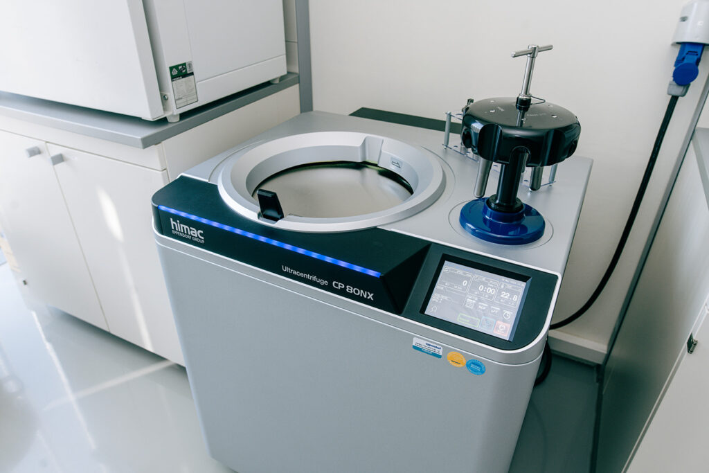
The Ultracentrifuge CP 80NX from Eppendorf Himac Technologies is equipped with a titanium P40ST swinging bucket rotor that can achieve a maximum relative centrifugal force (RCF) of 284,000 x g (40.000 rpm). Ultracentrifugation is employed to separate cellular organelles, to purify proteins or viral particles, and to isolate nucleic acids based on their density, size, and molecular weight. The rotor is ideal for density gradient centrifugation, which is typically applied for the isolation of extracellular vesicles. The rotor can be operated with 4 mL or 13 mL tubes and a maximum volume of 6 x 13 mL can be centrifuged per run.
Location: University for Continuing Education Krems, D3.09
Contact person: Susanne Krenn and Vladislav Semak, PhD
Agilent Bioanalyzer 2100
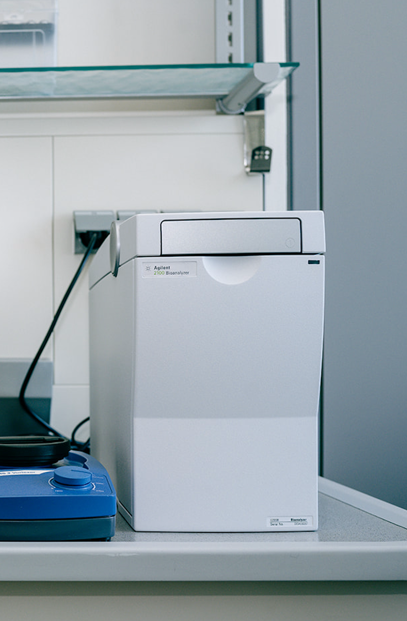
The Agilent 2100 Bioanalyzer system offers fast and reliable separation, sizing and quantification of proteins, DNA, and RNA by on-chip electrophoresis. The system enables high-sensitive detection in the picogram range for DNA, RNA, and protein analysis with minimal sample consumption (1 µL for nucleic acids and 4 µL for diluted proteins). Results are obtained in less than 30 minutes.
Material required:
- Agilent DNA kits (ideal for automated sizing and quantification of PCR fragments, restriction digests or fragmented DNA)
- Agilent RNA kits (for fast, easy and precise integrity checks and sample quantification)
- Agilent protein kits (for the assessment of protein concentration, identity, and purity in a wide variety of samples)
Location: University for Continuing Education Krems, D3.23
Contact person: Sabrina Summer, PhD
Parallelfermentersystem:
DG-System; Configurable DASGIP System
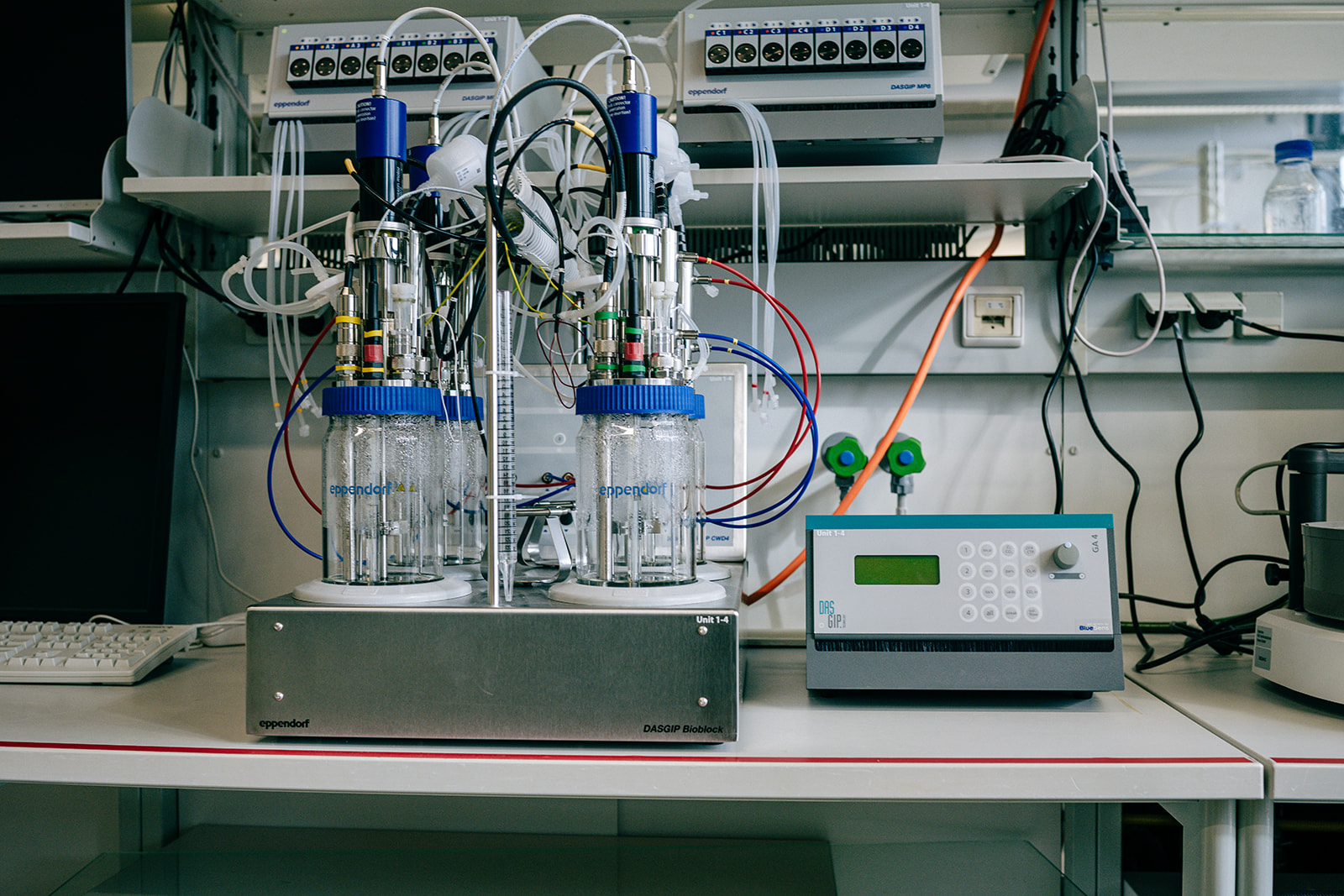
The DASGIP multi-fermenter system is an advanced bioprocessing platform designed for the cultivation of microbial and cell cultures. It allows for the simultaneous operation of multiple bioreactors, enabling high-throughput experimentation and optimization of bioproduction processes. The system is equipped with sophisticated control features for monitoring and adjusting crucial parameters such as temperature, pH, dissolved oxygen levels, and agitation speed. This level of control ensures precise and reproducible culture conditions, leading to consistent results and improved scalability. Additionally, the DASGIP multi-fermenter system offers flexibility in vessel configurations, accommodating various vessel sizes and types to suit different applications and experimental setups. Overall, it is a versatile and efficient tool for bioprocess development and optimization in research and industrial settings.
Technical details:
76 SR1000ODLS: Vessel size: 4 x 400 mL, Rushton Turbine: 2x with 4 blades
76DGCOND30: Offgas-condenser
76DGPHHMC220: pH-Sensor
76DGPOHVF220: DO-sensor
76DGLVL220: Antifoam-/Level-sensor
76DGTC4SC4B: Temperature control up to 99°C, Gas-mix module with up to 4 gases
76DGMX44H: Gasflow controlled by massflow-controler DASGIP MX4/4
76DGMP8: 16 flow controlled pumps
76DGIWF: Inline water filtration system
Location: IMC University of Applied Sciences Krems
Contact person: Prof.(FH) DI Dominik Schild
IXplore SPIN ScanR NoviSight
(confocal high content imaging microscope)
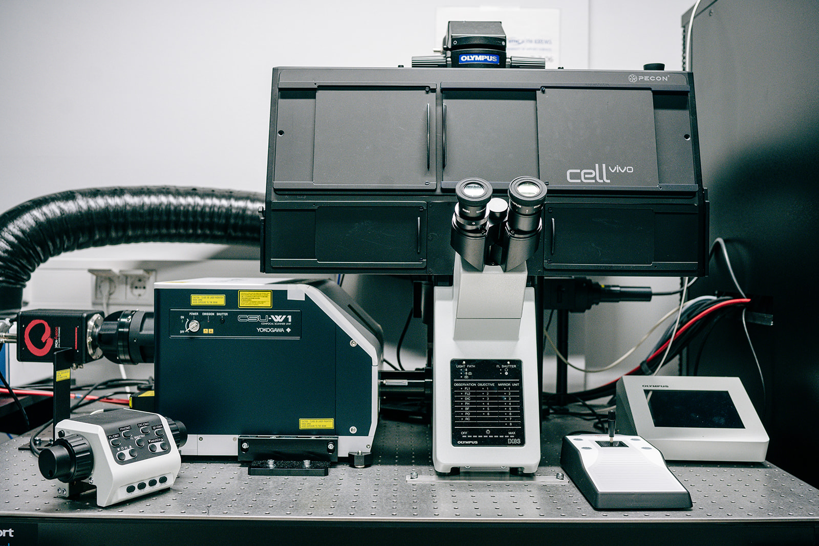
The microscope enables optimal imaging and precise 3D high content analysis of physiologically relevant 3D models such as reconstructed tumours, full skin equivalents or other 3D tissues.
The following applications are possible:
3D high-content analysis
Real object counting and morphological measurements in 3D.
Statistical analysis of microplate data (96 & 384 well plates).
Detection of rare cellular events.
Life Cell Imaging: Investigation of living cells using time-lapse microscopy.
Technical Details
Inverted motorised microscope stand for incident and transmitted light observation with 2 decks for optical module mounts and 1 slot for optional Z-drift compensator. Light intensity, observation, objective position, mirror turret position, beam path, fluorescence shutter position are shown on the integrated display. Beam path options for binocular tube and left side exit: 0:100, 50:50, 100:0. Motorised coaxial coarse and fine drive with nosepiece for raising/lowering, focusing stroke 9.5 mm. Maximum focussing speed: 3 mm/sec. Minimum step size: 0.01 μm. Optional accessory U-MCZ-1-2 for manual Z control. Compatible with transmitted light illumination IX3-ILL, IX3-RFA, IX3-RFAL, IX3-RFALFE reflected light illumination, IX3-SSU, BX3-SSU-1-2, IX3-SVR, IX3-SVL, IX2-SP, IX2-GS, GX-SVR, GX-SFR stages. Requires IX3-CBH control unit. Includes 6-position motorised IX3-D6REA nosepiece, touch panel control, jigs, power supply, cable clamps, control panel and nameplate.
Light source: IX3-LHLEDC-1-3 LED
White true colour LED light source for the IX83P2ZF, IX83P1ZF, IX73P2F, IX73P1F microscope stands with an intensity comparable to 30 W halogen light. Coupling to the microscope via the IX3-ILL transmission column. Light control via touch panel controller of IX83, U-MCZ or front control panel of IX73. Average LED service life 20,000 hours. U-LEDPS is required for use with IX73.
Laser:
- SD-COMB Laser Combiner
- IX-LAS405-50LXS laser 405 nm
- IX-LAS488-100LSS 488 nm laser
- IX-LAS561-100LSS laser system (561 nm)
- IX-LAS640-100LXS laser 640nm
Objectives:
Universal planapochromat objectives:
- UPLXAPO4X Objective (4X)
- UPLXAPO10X Objective (10X)
- UPLXAPO20X Objective (20X)
Universal apochromat water immersion objective:
- UAPON20XW340 NA 0.7 AA 0.35*BTS (20X)
- UAPON40XW340 NA1.15AA 0.25*BTS (40X)
- UPLSAPO60XW/1.2 objective (60X)
Universal Plan Semi-Apochromat, phase contrast objective:
- UCPLFLN20XPH (20X)
Filter: CSUW1P-DE09 Em. Filter 488/561
DAPI, FITC, Cy3, TexasRed, Cy5, Cy7
Software Modules:
- CS-S-DL-VF Deep Learning TruAI Solution
- CS-DI-V4.1 Dimension
- CS-DI-DT-V4.1 Dimension DT
- NS-ASW NoviSight main licence
Location: IMC University of Applied Sciences Krems / D2.05
Contact person: Prof.(FH) Mag. Dr. Christoph Wiesner
HP Workstation Z8 G4, Schrödinger Software
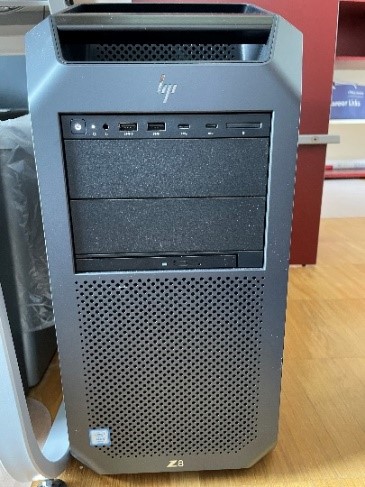
Advanced molecular modeling and 3D visualization of biomolecules
Location: IMC University of Applied Sciences Krems / D2.07
Contact person: Prof. (FH) Dr. Christian Klein
Stereo-Monitor 3D PluraView Impact 24″
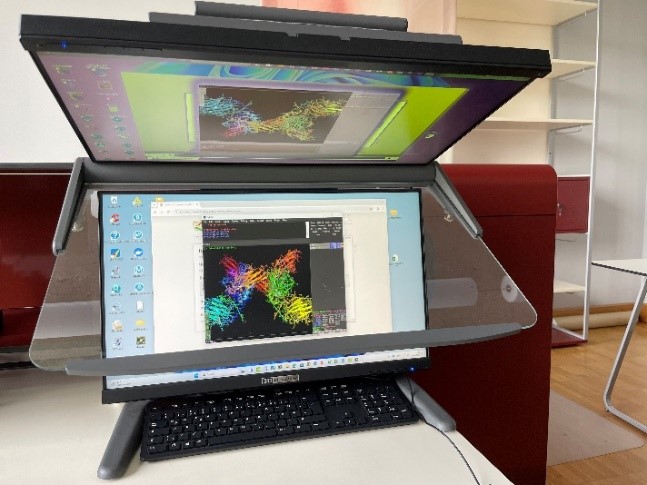
Full HD 144Hz B
Location: IMC University of Applied Sciences Krems / D2.07
Contact person: Prof. (FH) Dr. Christian Klein
MinON next generation sequencing device, Oxford Nanopore
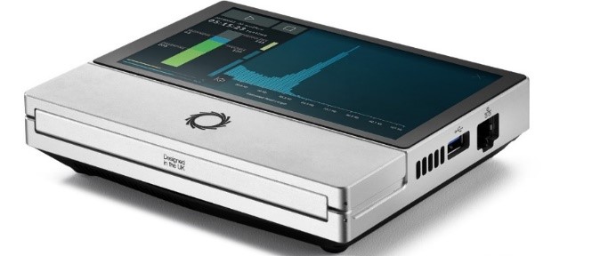
The Oxford Nanopore next generation sequencing device uses flow cells which contain nanopores embedded in an electro-resistant membrane. Each nanopore corresponds to its own electrode connected to a channel and sensor chip, which measures the electric current that flows through the nanopore. When a molecule passes through a nanopore, the current is disrupted to produce a characteristic ‘squiggle’. The squiggle is then decoded using basecalling algorithms to determine the DNA or RNA sequence in real time.
The device allows:
- Direct DNA/RNA sequencing; scalable
- Sequencing in real-time for rapid access to time critical information (e.g. pathogen identification), the generation of early sample insights and more control over the sequencing experiment
- Call base modifications simultaneously with nucleotide sequence
- Long reads to improve assembly and characterization of repetitive and difficult regions
- Applications include whole genome sequencing, targeted sequencing, metagenomics, epigenetics, epitranscriptomics.
Location: IMC University of Applied Sciences Krems / D.E.01
Contact person: Prof. (FH) Priv.-Doz. Dr. Reinhard Klein
Lonza 4D Nucleofector

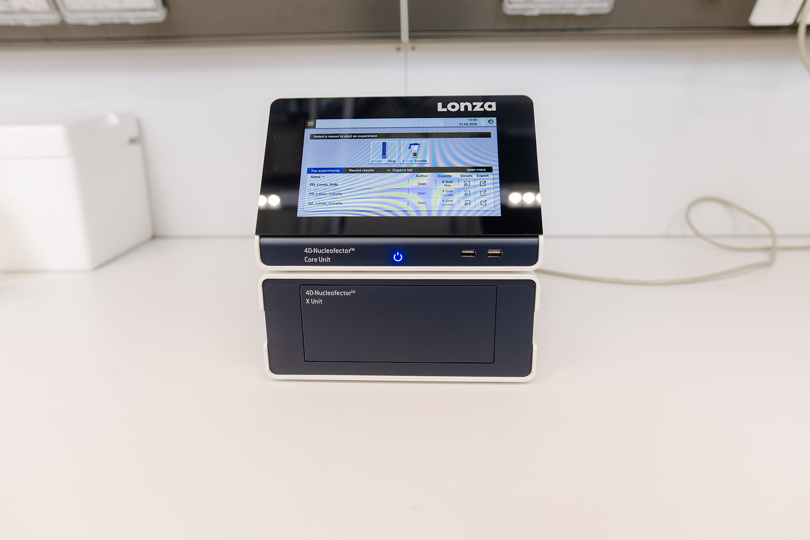
The 4D-Nucleofector System with Y Unit is an electroporation platform designed for efficient transfection of primary cells, stem cells, and hard-to-transfect cell lines. Using optimized electrical parameters, it enables direct delivery of DNA, RNA, or proteins into the nucleus or cytoplasm with high efficiency and reproducibility, while maintaining cell viability.
TThe Y Unit supports transfection in 16-well strips, making it suitable for medium-throughput experiments and comparative studies. It provides precise control over electroporation conditions, enabling standardized workflows and reproducible results across multiple cell types.
Applications include genetic modification of primary cells, transient or stable expression studies, CRISPR/Cas9 genome editing, and RNA interference experiments. The 4D-Nucleofector with Y Unit offers a versatile platform for researchers working with challenging cell systems in molecular and cellular biology.
Contact person: rajeshwari.meli@kl.ac.at
Tecan Spark Plate Reader

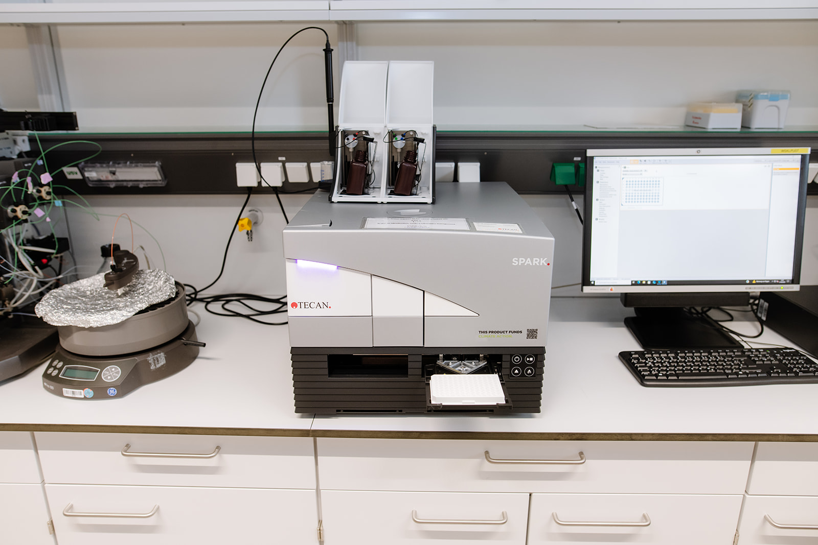
The Tecan Spark Plate Reader is a versatile detection system for fluorescence, luminescence, and absorbance measurements. In addition to standard microplate formats, it includes an injector system that enables real-time assay setup by adding reagents or substrates directly to the plate during or immediately before measurements. A cuvette port is also available for flexible sample handling outside of plates. The NanoQuant Plate allows precise quantification of nucleic acids and proteins from very small sample volumes.
The instrument supports both endpoint and kinetic assays, making it suitable for a broad range of experimental designs. Typical applications include enzyme activity measurements, nucleic acid and protein quantification, reporter gene assays, cell-based assays, and luminescence-based detection. With its combination of sensitivity, flexibility, and throughput, the Spark Plate Reader is well suited for research in molecular biology, biochemistry, and cellular studies.
The system is equipped with the following filters:
320/25, 340/20, 485/20, 510/10, 535/25, 560/20, 595/35, 620/10, 620/20, 665/8.5, 680/30
Location: Karl Landsteiner University of Health Sciences
Contact persons: Prof. Dr. Gerald Obermair; Prof. Dr. Dagmar Stoiber-Sakaguchi
ÄKTA Pure 25L

The ÄKTA Pure system is a modular chromatography platform designed for protein purification and characterization. Equipped with an F9-C fraction collector and an S9 sample pump, it supports flexible workflows from sample loading to automated fraction collection. The system is suitable for both routine purification and method development, offering precise flow control and monitoring for reproducible results.
Proteins are detected via UV absorbance at 280nm and integrated conductivity and pH monitoring enable detailed process control.
Available columns include:
- HisTrap FF Crude (5 × 1 ml): Fast purification of histidine-tagged proteins directly from unclarified lysates; compatible with reducing/denaturing agents and detergents.
- GSTrap FF (1 × 5 ml): Purification of GST fusion proteins with capacity for higher flow rates at wash and elution.
- HiLoad 26/600 Superdex 75 pg: High-resolution preparative purification of proteins and peptides (Mr 3,000–70,000).
- Superdex 200 Increase 10/300 GL: Analytical gel filtration for proteins in the Mr 10,000–600,000 range.
This configuration enables rapid affinity, ion-exchange, and size-exclusion chromatography, making the ÄKTA Pure system suitable for diverse protein purification tasks in structural biology, biochemistry, and molecular biology research.
Location: Karl Landsteiner University of Health Sciences
Contact persons: Prof. Dr. Gerald Obermair ; Dr. Manuel Hessenberger
